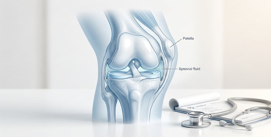




Talonavicular arthritis is wear or inflammation in the joint at the top of the arch, where the talus meets the navicular bone. It is one of the most common causes of midfoot pain, and also one of the most frequently missed, because the first symptom is usually nothing more dramatic than ten or twenty stiff steps in the morning. Left alone, the cartilage in the joint thins, bone spurs form along the rim, and shoes that fit perfectly for years start to feel tight across the instep.
This guide covers where the joint sits, what wears it down, the early signs worth acting on, how talonavicular arthritis is diagnosed, and what the options are, from shoe changes and bracing through to talonavicular fusion.
What is the Talonavicular Arthritis?
Talonavicular arthritis is the loss of healthy cartilage in the talonavicular joint, the ball-and-socket joint on the inner side of the foot where the rounded head of the talus sits against the cup of the navicular bone. Cartilage is the smooth, low-friction surface that lets the two bones glide across one another. As it thins, bone begins to contact bone, the joint becomes inflamed, and the body responds by laying down extra bone around the rim in the form of spurs, or osteophytes.
The condition arrives by several different routes. Most cases are degenerative, meaning the cartilage has simply worn under decades of load. Some follow an injury years earlier, which is called post-traumatic arthritis. Others are driven by an inflammatory disease such as rheumatoid arthritis, which attacks the joint lining directly. And a large group is mechanical in origin, where a flattening arch changes how force passes through the joint and concentrates it along one edge.
Because the talonavicular joint is one half of the Chopart joint, the dividing line between the hindfoot and the midfoot, arthritis here affects far more than a single point on the foot. It limits the rotation the foot needs to adapt to uneven ground, which is why many people notice the problem on grass or gravel long before they notice it on pavement.
Causes of Talonavicular Arthritis
Talonavicular arthritis isn’t just an old person’s disease. It’s something that you can get regardless of your age. It happens when the cartilage that cushions your bones and joints becomes too thin – and at that point, bones and foot joints rubbing against each other causes progressive damage.
But, why do some people get talonavicular arthritis? This leads us to the causes. There isn’t a single fixed cause and it can differ from person to person. Yet, we can take a closer look at a few common reasons why people get this type of arthritis.
- If you’ve got osteoarthritis, you’re at risk.
- Inflammatory arthritis (like Rheumatoid arthritis) can also affect your talonavicular joint.
- A foot injury can also cause the symptoms.
Injury is a very common cause. In fact, the American Academy of Orthopedic Surgeons suggests that you’re about 700% more likely to get arthritis in a joint that you’ve hurt – compared to a healthy joint, of course.

Talonavicular Arthritis Symptoms
Knowing the symptoms of talonavicular arthritis helps you get a diagnosis early on. So, what should you look out for? Here are some symptoms you may notice:
- Pain and swelling
- Stiffness
- Worse pain if you put weight on your foot
- Stiffness and pain are worse in the morning
- Tenderness
If you’ve got these Arthritis symptoms, note that it can mean other things too. Getting your doctor to take a look at your foot and run some tests helps to find the cause. If it’s talonavicular arthritis, they’ll discuss the treatment options with you.
The Impact of Talonavicular Arthritis on Foot during Walking
If you’ve got talonavicular arthritis, then it’s going to take a toll on your walking capabilities. It’s important to understand why this is.
When you walk, you’re actually relying on your talonavicular joint to have flexibility in your feet. See, as you take a step, your ankle might be moving upward and downward – but it’s not the same for this joint. The joint actually moves to the sides. It’s critical to ensure you can walk on different types of surfaces. And when you’ve got this type of arthritis, it can hinder the joint’s ability to move properly as you walk.
Diagnosis of Talonavicular Arthritis
Diagnosis rests on a hands-on examination first and imaging second. The midfoot contains a dozen small joints packed into a few inches, so the examination is largely about narrowing down which one is generating the pain.
- The examination. A foot and ankle specialist will press directly over the talonavicular joint, on the inner side of the foot just forward of the ankle, looking for tenderness over the joint line itself rather than over the tendon that runs beside it. They will then hold the heel still and rotate the forefoot inward and outward. Reproducing pain with that rotation while ankle movement stays comfortable is one of the more telling findings, because it isolates the Chopart joint from the ankle above it. Expect them also to watch you walk, check whether the arch flattens as you bear weight, ask you to perform a single-leg heel raise to test the tibialis posterior tendon, and compare both feet, since arthritis driven by inflammatory disease usually shows up on both sides.
- Weight-bearing X-rays. These are the first imaging step and they need to be taken standing, not lying down. A joint can look almost normal unloaded and then show clear narrowing once body weight passes through it. The views typically ordered are a standing anteroposterior and lateral of the foot. Radiologists look for four things: narrowing of the space between the talus and the navicular bone, osteophytes along the top of the joint, hardening of the bone just under the joint surface, and any sag in the arch measured by the angle between the talus and the first metatarsal.
- CT scan. A CT gives a far more precise picture of how much cartilage is left and which neighboring joints are involved. It is ordered most often when surgery is being considered, because it shows whether the naviculocuneiform and calcaneocuboid joints also need addressing. Weight-bearing CT, where available, is becoming the preferred option because it captures the alignment of the foot under load in three dimensions.
- MRI. An MRI is less about the arthritis and more about everything around it. It shows the tibialis posterior tendon, the spring ligament and the talonavicular ligament, and it will pick up a stress fracture of the navicular bone or early bone marrow changes that X-rays miss entirely. It is the test of choice when the pain does not match the X-ray findings.
- Diagnostic injection. Where two or three midfoot joints look abnormal on imaging and it is unclear which one hurts, a surgeon may inject a small amount of local anesthetic into the talonavicular joint under ultrasound or fluoroscopic guidance. If the pain settles for a few hours, that joint is confirmed as the source. This matters a great deal before fusion surgery, because fusing the wrong joint leaves the pain behind.
- Blood work. If both feet are affected, if the stiffness lasts well beyond half an hour each morning, or if other joints are involved, your physician may order inflammatory markers and rheumatoid testing to look for an underlying inflammatory arthritis rather than simple wear.
- What else it could be. Several conditions sit close enough to be confused with talonavicular arthritis: tibialis posterior tendon dysfunction, which produces pain slightly lower and behind the navicular bone; a navicular stress fracture, more common in runners and typically far more focal; an accessory navicular, an extra bone present from birth that becomes painful in adolescence; a tarsal coalition; and gout, which arrives suddenly with redness and warmth rather than building over years. Each is distinguished by a combination of where the tenderness sits and what the imaging shows.
Complications of Talonavicular Arthritis
The biggest problem with talonavicular arthritis is the fact that it’s going to affect your ability to walk. Let’s face it – we rely on walking every day. So, when your talonavicular joint gets damaged, you may have pain when you walk. And as you put pressure on your feet, the problem can get worse.
Over time, this arthritis can cause deformities. This means the actual structure of your joints and the foot itself can change. When this happens, it’s going to make walking even harder. Eventually, it’s something that can actually make you reliant on things like a walking crane. And, in severe cases, you might not be able to walk anymore. This means you may need to start using things like a wheelchair.
Talonavicular Joint Arthritis Treatment Options
You’ve been diagnosed with arthritis in your talonavicular joint – now what? Well, treatment depends on your situation. A few imaging tests helps your doctor see how serious the problem is. This way, they can give you personalized treatment.
Start with home remedies – this is usually the initial treatment. Resting is important. Don’t put too much pressure on your feet. At the same time, you’ll still need to do some light exercises to keep up with your fitness. If you’re overweight, you might want to consider losing excess weight. This way, you’ll also lessen the pressure you put on your feet when you stand up and walk.
What type of shoes do you wear? The right footwear can actually make a big difference. Wear shoes that stabilize your feet and ankles. You can talk to your doctor about orthotics. Sometimes, a brace can help too.
We’ll also need to consider medical treatments. There are anti-inflammatories and pain relievers that help with the symptoms you experience. Sometimes, steroid injections can be helpful too. They help with swelling, but it’s also just a short-term option.
For a more natural approach, look at a Flexoplex review. It’s a supplement that you take daily, and it helps with joint health. Plus, it can reduce some of the symptoms you experience.
If your arthritis is bad, then surgery is something to consider. Both reconstruction and fusion surgery can help. If there’s a lot of damage in the talonavicular joint, then reconstruction helps to build the joint back up. It’s also possible to work on the cartilage that was damaged – this can help to offer more support for the joint and reduce bone-on-bone rubbing. Fusion surgery, on the other hand, works by fusing some joints together. This means the joints won’t move like usual – but it can help with talonavicular arthritis.
Fusion surgery uses staples, pins, or screws to fuse the joints together. You’ll be wearing a cast for about 12 weeks. Rehabilitation is also something that you’ll need if you opt for this surgery. It’s effective, though. A study in the Journal of Foot & Ankle Surgery looked at how the procedure affected people – and everyone in the follow-up said they had improvements in their symptoms.

Nonsurgical Treatment for Talonavicular Arthritis
Most people with talonavicular arthritis never need surgery. The cartilage that has gone will not come back, but the joint can often be made comfortable by changing how much it has to move and how much load it has to carry. That is the whole logic of nonsurgical care here, and it is why footwear does more work than any other single measure.
- Stiffen the sole. A rigid or semi-rigid sole stops the midfoot from bending through the painful part of its range. This is the reason so many people with midfoot arthritis discover that their hiking boots feel better than their running shoes. A carbon fiber or spring steel insert slipped into an existing shoe achieves the same thing cheaply, and a cobbler can add a rocker sole, a curved profile that lets the foot roll forward without flexing at the arch. For many people this combination alone changes the day.
- Support the arch. Because a flattening arch concentrates load on one edge of the joint, a custom orthotic with a deep heel cup and firm medial arch support helps distribute it more evenly. Over-the-counter arch supports are worth a try first; a podiatrist can take a cast for a custom device if the off-the-shelf version helps but not enough.
- Bracing for more advanced cases. Where the arch has collapsed significantly or pain persists in good shoes, a lace-up ankle brace or a custom Arizona-style brace, which is a leather gauntlet that wraps the ankle and hindfoot, restricts the motion that provokes the pain. These are bulkier and take some getting used to, but they are often what keeps someone on their feet at work.
- Adjust activity rather than stop it. Swapping running for cycling, swimming, rowing or an elliptical trainer keeps fitness and leg strength up while taking repeated impact out of the midfoot. Walking on pavement is generally kinder than walking on sand, grass or gravel, because flat ground asks less rotation of the joint. Reducing time spent standing still, which loads the joint without the muscle pumping that movement provides, tends to help more than people expect.
- Physical therapy. A therapist will usually work on three things: calf flexibility, because a tight calf forces the midfoot to make up the difference; tibialis posterior and intrinsic foot strength, which supports the arch dynamically; and balance work, which reduces the small corrective movements that aggravate the joint. Pain that lingers more than a day after a session means the dose was too high, not that therapy is the wrong idea.
- Weight management. Every pound of body weight translates into several pounds of force through the midfoot at each step. Where weight loss is appropriate, it is one of the more effective levers available, and it compounds with everything else on this list.
- Anti-inflammatory medication. Oral nonsteroidal anti-inflammatory drugs can make a meaningful difference to pain and swelling during a flare, and topical versions applied over the joint deliver the drug locally with less systemic exposure, which matters for older adults and anyone with stomach, kidney or cardiac concerns. Both are short-term tools rather than long-term plans, and dosing should be set by your physician or pharmacist.
- Corticosteroid injection. An image-guided steroid injection into the joint can quiet things down for weeks to months. It serves two purposes at once: it buys comfort, and the degree of response tells the surgeon how much of the pain is actually coming from this joint. Injections are generally limited in number because repeated steroid exposure is unhelpful for the remaining cartilage and the surrounding soft tissue.
- Supplements as part of a broader routine. Ingredients such as glucosamine, chondroitin, collagen peptides and omega-3 fatty acids appear in products marketed to support joint comfort and mobility, and formulas such as Flexoplex combine several of them. Evidence in midfoot arthritis specifically is limited, so they are best thought of as one element alongside footwear, bracing, therapy and weight management rather than a replacement for any of them. Check with your physician or pharmacist first, particularly if you take blood thinners or have a shellfish allergy.
When to See a Foot and Ankle Specialist
Book an appointment if midfoot pain has lasted more than six weeks despite better shoes and reduced activity, if you cannot get through a normal working day on your feet, if the arch is visibly flattening or one foot now looks different from the other, if a bony bump on top of the foot is rubbing in every shoe you own, or if there is redness, warmth or swelling that came on over hours rather than months, which needs assessment promptly. Sudden severe pain after a twist or a fall should be seen straight away to rule out a fracture.
Frequently Asked Questions
What part of the foot is the talonavicular joint?
This joint helps with circular motion. It’s also a joint that makes sure you can move your foot outward and inward. Part of the ankle joint, called the talus, and some of the bone in your feet (at the front area of your navicular) form the talonavicular joint.
What is the function of the talonavicular ligament?
It’s a ligament that’s covered by extensor tendons. It acts as a connector between the navicular bone’s dorsal surface and the talus’s neck. You may notice that some people call the ligament the superior astragalonavicular ligament or the dorsal talonavicular ligament.
Can foot arthritis cause swelling?
Yes, if you’ve got foot arthritis, then you’ll likely notice some swelling. This condition doesn’t only cause pain and swelling, but will also make your feet feel stiff.
What is the best exercise for arthritic feet?
Toe stretches, Achilles stretches, and sand walking are some good options. You don’t want to overdo it, so choose exercises that won’t put a lot of stress on your feet. Other exercises include swimming and riding a bike.
Key Takeaways
If you’ve got talonavicular arthritis, then it’s really important to take action before it affects your ability to walk properly. You’ve got to get a diagnosis first, so the process starts with a visit to your doctor’s office. Luckily, there are some things you can do, like wearing the right footwear and resting. Some medications can help, and, if it’s really bad, surgery might also be a good option, in which case a surgeon can address the damage done to your foot joints.
References
https://www.jfas.org/article/S1067-2516(10)00294-2/abstract
https://pmc.ncbi.nlm.nih.gov/articles/PMC5620404/